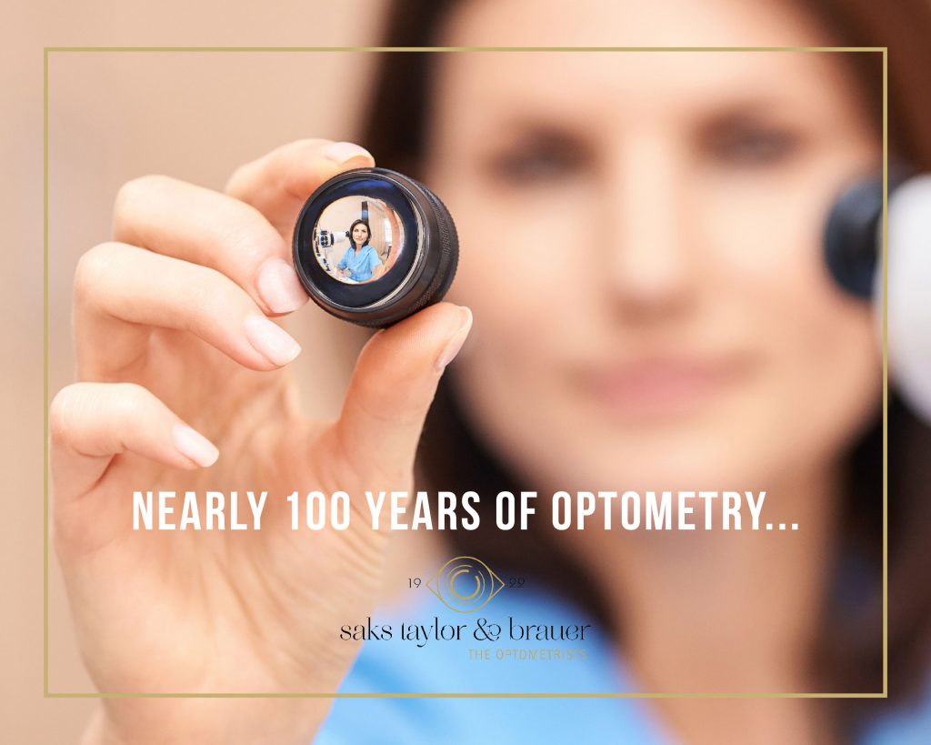
Saks, Taylor and Brauer optometrists are almost one hundred years old! A lot has changed since 1922 when Julius Louis Saks set up the practice as an optician near the southwest corner of Paul Kruger and Pretorius Street.
Over the last century, Saks Taylor and Brauer optometrists have invested in the latest and best technology. By investing in technology, we invest in your eye care and eye health.
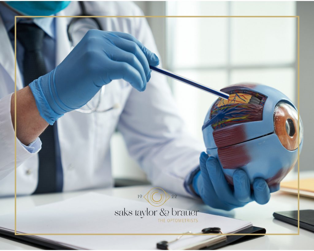
We have different testing equipment, making sure we can truly offer a comprehensive eye test.
Let us look at these different tests and start with the test that all the patients will always remember, the one that puffs air into the eye causing a startled reaction: the tonometer.
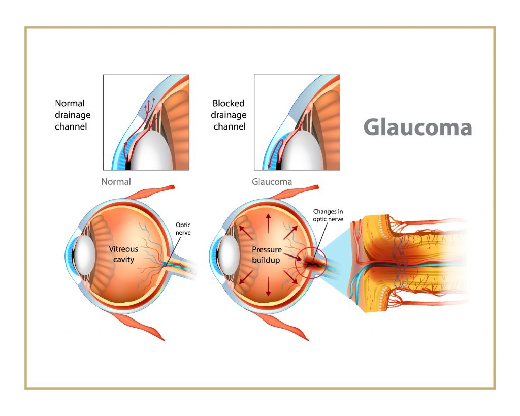
This measures your eye pressure, and it is especially important that your pressure does not go too high as it can cause damage to the optic nerve. This condition is called Glaucoma. This is a treatable condition and early detection is crucial to maintain optimal vision, because if left untreated it can lead to irreversible vision loss and blindness.
We perform Optical Coherence Tomography which is a very specialized scan that uses light waves to obtain cross-sectional scans of the retina. It is non-invasive and provides an image like a CT scan that allows us look at each retinal layer. This scan will allow us to detect a disease long before the patient is even aware that there is a problem. Early detection and treatment may reduce the risks of vision loss and disease progression.
Next, the corneal topographer is a computerized test that 3D maps the shape and curve of your cornea, the outer surface of the eye. These details are used to diagnose and monitor eye conditions. It is also used in the fitting process of specialized contact lenses like rigid gas permeable and scleral lenses.
At Brooklyn we can also do Meibography by using the oculus keratographer. A series of photographs are taken of the meibomian glands, non-invasive tear break-up time and tear meniscus height measurements and evaluation of the lipid layer are done to diagnose the type of dry eye and to monitor dry eye disease. More specific dry eye drops can then be prescribed pending the outcome and in office treatment can be arranged in severe cases with the blephasteam to treat and assist dry eye disease.
The next test is the visual field test which is a method of measuring an individual’s entire scope of vision, their central and peripheral vision. Visual field testing maps the visual field of each eye individually and can detect blind spots as well as more subtle areas of dim vision. Visual field testing is also used to detect signs of glaucoma damage to the optic nerve. It is also useful for detecting central and peripheral retinal diseases of the retina and eyelid conditions such as drooping (ptosis) and conditions affecting the visual pathways from the optic nerve to the area of the brain where this information is processed into vision.
The fundus camera is a specialized low power microscope with an attached camera that takes a photograph of the interior of your eye through the pupil. Fundus photographs are visual records which document the current ophthalmoscopic appearance of a patient’s retina. One picture is worth, in this instance, a thousand words in the optometrist’s notes. This is used to further study the retina and to identify retinal changes on follow-up examinations.
We know that for the best vision, optimal eye health needs to be maintained and early detection and diagnosis of changes is crucial. That is why we have invested in the different testing equipment over the last century, so that we can continue to monitor your eye health and take the best possible care of your eyes for the next one hundred years to come!